Literature References
Covalent Inhibition
The article discusses recent innovations in covalent drug discovery, covering various aspects such as the design principles, mechanisms of action, and applications of covalent drugs. It emphasized how advancements in chemical biology and medicinal chemistry have enabled
the development of covalent drugs that target specific disease mechanisms with enhanced potency and selectivity. The review also highlights the potential of covalent drugs in treating challenging diseases in the future.
The article explores the emergence and increasing importance of targeted covalent inhibitors in drug discovery. It discusses how these inhibitors are designed to form durable bonds with specific target proteins, leading to enhanced potency and selectivity compared to traditional non-covalent inhibitors. The review covers key principles in the design and optimization of targeted covalent inhibitors, as well as their applications across various disease areas.
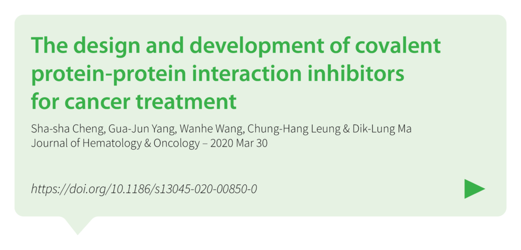
The article addresses the challenges associated with targeting PPIs, which are crucial for cellular signaling and often implicated in cancer progression. It discusses various approaches and techniques employed in the design of covalent PPI inhibitors, emphasizing the need for specificity and potency. Case studies and examples are provided in preclinical and clinical settings, highlighting their potential in overcoming resistance mechanisms and enhancing treatment efficacy in cancer.
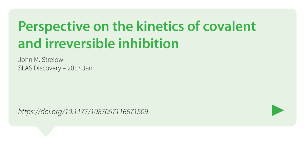
This perspective article discusses fundamental concepts such as the mechanisms of covalent binding between drugs and their targets, as well as factors influencing the kinetics of these interactions. The author explores how understanding the kinetic properties of covalent and irreversible inhibitors can inform drug design and optimization processes. The article also addresses the implications of kinetic parameters on efficacy, selectivity, and safety profiles of covalent drugs.
The article discusses the advantages of covalent drugs regarding enhancing potency and selectivity. It highlights the challenges in designing covalent drugs, such as ensuring specificity and minimizing off-target effects to minimize potential toxicity. The review also covers various strategies for optimizing covalent drug candidates, including medicinal chemistry approaches and the use of advanced screening techniques.
Diabetes and Beta Cell Function
Type 2 diabetes-a matter of beta-cell life and death?
Christopher J. Rhodes
Science – 2005 Jan 21, 307(5708):380-4. doi: 10.1126/science.1104345. PMID: 15662003.
https://pubmed.ncbi.nlm.nih.gov/15662003/
The article explores the pivotal role of beta-cell dysfunction and death in the pathophysiology of type 2 diabetes mellitus (T2DM). It discusses how the progressive loss of beta-cell function and mass contributes to the inability to maintain normal blood glucose levels in individuals with T2DM, highlighting factors such as genetic predisposition, lifestyle choices, and environmental factors that influence beta-cell health and function. The review suggests that strategies aimed at preserving beta-cell mass and function could hold promise for preventing and managing T2DM.
Diabetes Invest_Beta-cell failure in diabetes – Common susceptibility and mechanisms shared between type 1 and type 2 diabetes 1
Hiroshi Ikegami, Naru Babaya, and Shinsuke Noso
Journal of Diabetes Investigation – 2021 Sep; 12(9): 1526–1539. Published online 2021 Jun 16. doi: 10.1111/jdi.13576
https://www.ncbi.nlm.nih.gov/pmc/articles/PMC8409822/
The article explores the shared susceptibility and underlying mechanisms of beta-cell dysfunction in both T1DM and T2DM. It discusses how genetic predisposition, autoimmune responses, and environmental factors contribute to the loss of beta-cell function in both forms of diabetes. The review emphasizes future research directions aimed at addressing beta-cell failure as a key strategy in managing and potentially preventing both T1DM and T2DM.
Not control but conquest – Strategies for the remission of Type 2 diabetes mellitus
Jinyoung Kim, Hyuk-Sang Kwon
Diabetes and Metabolism Journal – 2022 Mar;46(2):165-180. doi: 10.4093/dmj.2021.0377.
https://pubmed.ncbi.nlm.nih.gov/35385632/
The article explores strategies aimed at achieving remission rather than mere control of T2DM. It discusses various approaches including lifestyle modifications, pharmacotherapy, and bariatric surgery, which have shown potential in achieving sustained remission of T2DM. It also emphasizes personalized treatment plans tailored to individual patient characteristics such as age, duration of diabetes, and comorbidities. The review also covers emerging therapies such as GLP-1 receptor agonists and SGLT-2 inhibitors, which have demonstrated efficacy in improving beta-cell function and insulin sensitivity.
Increased beta-cell proliferation before immune cell invasion prevents progression of Type 1 diabetes
Dirice E et al., 2019 Nature Metabolism
Nat Metab – 2019 May; 1(5): 509–518. Published online 2019 May 6. doi: 10.1038/s42255-019-0061-8
https://www.ncbi.nlm.nih.gov/pmc/articles/PMC6696912/
The study explores how enhanced beta-cell replication prior to immune cell infiltration can mitigate the development of T1D. The researchers utilize mouse models and human pancreatic tissue to demonstrate that beta-cells exhibit increased proliferation in response to specific conditions, which leads to improved glucose tolerance and delays in T1D onset. The study underscores the potential therapeutic implications of promoting beta-cell proliferation as a strategy to prevent or delay the progression of T1D.
Importance of beta cell mass for glycaemic control in people with Type 1 diabetes
Diabetologia, 2023 Feb; 66(2):367-375. doi: 10.1007/s00125-022-05830-2. Epub 2022 Nov 17.
https://pubmed.ncbi.nlm.nih.gov/36394644/
The article discusses how the preservation or restoration of beta-cell mass is crucial for achieving stable blood glucose levels and minimizing complications associated with T1D. It reviewed current research and clinical evidence highlighting that even small residual amounts of beta-cell function can significantly improve glycemic outcomes and reduce the risk of hypoglycemia.
Postprandial C-Peptide to Glucose Ratio as a Marker of β Cell Function: Implication for the Management of Type 2 Diabetes
International Journal of Molecular Science – 2016 May 17; 17(5):744. doi: 10.3390/ijms17050744. PMID: 27196896; PMCID: PMC4881566.
https://pubmed.ncbi.nlm.nih.gov/27196896/
The article examines the utility of the postprandial C-peptide to glucose ratio as an indicator of beta-cell function in individuals with T2D and discusses how this ratio reflects the insulin secretion relative to glucose levels after meals, providing insights into beta-cell health and function beyond fasting measurements. The review suggests that monitoring postprandial C-peptide to glucose ratios could improve the management of T2D by assessing beta-cell responsiveness and predicting treatment outcomes as a valuable tool in personalized diabetes care.
Remission of human Type 2 diabetes requires decrease on liver and pancreas fat content, but is dependent upon capacity for beta cell recovery
Cell Metabolism, 2018 Oct 2; 28(4):547-556.e3. doi: 10.1016/j.cmet.2018.07.003.
https://pubmed.ncbi.nlm.nih.gov/30078554/
The study explores how reducing fat accumulation in the liver and pancreas is critical for T2D remission. It highlights that while weight loss and fat reduction are crucial, sustained remission also depends on the capacity of beta cells to recover and improve insulin secretion. The findings underscore the complex interplay between metabolic factors and beta-cell health in achieving and maintaining T2D remission, suggesting comprehensive approaches that address both fat accumulation and beta-cell recovery are essential for long-term management of the disease.
Intervention with therapeutic agents, Understanding the path to remission in Type 2 Diabetes: Part 1
Shuai Hao et al.
Endocrinology Metabolism Clinics of North America – 2023 Mar; 52(1):27-38. doi: 10.1016/j.ecl.2022.07.003. Epub 2022 Nov 14.
https://pubmed.ncbi.nlm.nih.gov/36754495/
The article explores various therapeutic interventions aimed at achieving remission in T2D, including strategies such as lifestyle modifications, pharmacotherapy, and bariatric surgery. It reviewed current research on the mechanisms underlying these interventions, including their effects on insulin sensitivity, beta-cell function, and metabolic pathways. The article emphasizes the importance of personalized treatment approaches tailored to individual patient characteristics and disease progression.
Intervention with therapeutic agents, Understanding the path to remission in Type 2 diabetes – Part 2
Shuai Hao et al.
Endocrinology Metabolism Clinics of North America – 2023 Mar; 52(1):39-47. doi: 10.1016/j.ecl.2022.07.004. Epub 2022 Nov 18.
https://pubmed.ncbi.nlm.nih.gov/36754496/
The article continues to explore therapeutic interventions aimed at achieving remission in T2D by focusing on pharmacological agents and their mechanisms of action in improving glycemic control and potentially inducing remission. They discuss the efficacy and safety profiles of various medications, including insulin sensitizers like metformin, GLP-1 receptor agonists, SGLT-2 inhibitors, and other emerging therapies. The review highlights clinical evidence and guidelines for using these agents, emphasizing their roles in reducing cardiovascular risks and enhancing overall metabolic health in T2D patients.
Beta Cell Proliferation
Pancreatic β-cell proliferation in obesity
Linnemann AK, Baan M, Davis DB
Advances in Nutrition – 2014 May 14, 5(3):278-88. doi: 10.3945/an.113.005488. PMID: 24829474; PMCID: PMC4013180.
https://pubmed.ncbi.nlm.nih.gov/24829474/
The review explores the phenomenon of pancreatic beta-cell proliferation in the context of obesity. The authors discuss how obesity, characterized by excess adiposity, insulin resistance, and metabolic dysregulation, impacts beta-cell function and mass. They examine mechanisms through which obesity-related factors such as nutrient excess, inflammation, and insulin resistance influence beta-cell proliferation and survival. The review suggests that understanding these mechanisms is crucial for developing strategies to preserve beta-cell function and prevent diabetes in the context of obesity-related metabolic disorders.
A morphological study of the endocrine pancreas in human pregnancy
F.A. Van Assche FA, L. Aerts L, F. De Prins
British Journal of Obstet Gynaecology – 1978 Nov, 85(11):818-20. doi: 10.1111/j.1471-0528.1978.tb15835.x. PMID: 363135.
https://pubmed.ncbi.nlm.nih.gov/363135/
The study investigates changes in the structure of the endocrine pancreas during pregnancy. The authors examine pancreatic tissue samples from pregnant women to analyze alterations in the size, distribution, and function of pancreatic islet cells, which are responsible for insulin production. The study underscores the physiological adjustments of the pancreas during gestation, highlighting the relevance for understanding metabolic changes and potential implications for maternal health and fetal development.
Beta cell adaptation to pregnancy requires prolactin action on both beta and non-beta cells
Shrivastava V, Lee M, Lee D, Pretorius M, Radford B, Makkar G, Huang C.
Scientific Reports – 2021 May 14, 11(1):10372. doi: 10.1038/s41598-021-89745-9. PMID: 33990661; PMCID: PMC8121891.
https://pubmed.ncbi.nlm.nih.gov/33990661
The study explores how prolactin, traditionally associated with lactation, influences beta-cell function and proliferation. The authors utilize experimental models to demonstrate that prolactin acts on both beta cells and non-beta cells within the pancreas, enhancing beta-cell mass and insulin secretion capacity. The study highlights that these adaptations are crucial for meeting increased insulin demands during pregnancy, ensuring maternal glucose homeostasis.
Prolactin-regulated Pbk is involved in pregnancy-induced β-cell proliferation in mice
Cao, Y., Feng, Z., He, X., Zhang, X., Xing, B., Wu, Y., Hojnacki, T., Katona, B. W., Ma, J., Zhan, X., & Hua, X. (2022)
Journal of Endocrinology, 252(2), 107-123.
https://doi.org/10.1530/JOE-21-0114
The study explores how prolactin signaling activates PBK, a key mediator in promoting the expansion of beta-cell mass in response to the metabolic demands of pregnancy. Using mouse models, they demonstrate that PBK plays a critical role in enhancing beta-cell proliferation, thereby facilitating increased insulin production necessary for maternal glucose regulation during gestation.
Expansion of β-cell mass in response to pregnancy
Sebastian Rieck and Klaus H. Kaestner
Trends in Endocrinology and Metabolism – 2010, Mar 21, (3):151-8. doi: 10.1016/j.tem.2009.11.001. Epub 2009 Dec 16.
https://pubmed.ncbi.nlm.nih.gov/20015659/
The review article discusses how pregnancy induces profound changes in pancreatic beta-cells, leading to their proliferation and expansion. It examines molecular pathways and hormonal factors involved in regulating beta-cell growth, including the roles of prolactin, placental lactogens, and insulin-like growth factors. The review highlights the adaptive responses of beta-cells to the increased metabolic demands during gestation, emphasizing the importance of these mechanisms in maintaining maternal glucose homeostasis.
Beta-cell compensation and gestational diabetes
Taofeek O Usman, Goma Chhetri, Hsuan Yeh, H Henry Dong
Journal of Biological Chemistry – 2023, Dec, 299(12):105405. doi: 10.1016/j.jbc.2023.105405. Epub 2023 Oct 29.
https://pubmed.ncbi.nlm.nih.gov/38229396/
The article investigates the mechanisms of beta-cell compensation in the context of gestational diabetes mellitus (GDM). The authors explore how pregnancy induces adaptations in beta-cell function to meet increased insulin demands, emphasizing the role of compensatory mechanisms when insulin sensitivity is impaired. The review addresses the implications of these adaptations for the development and management of GDM, highlighting potential targets for therapeutic interventions aimed at preserving beta-cell function and improving maternal and fetal outcomes.
Serum from pregnant donors induces human beta cell proliferation and insulin secretion
Sylvester-Armstrong KR et al.
bioRxiv – 2023, Apr 17, 2023.04.17.537214. doi: 10.1101/2023.04.17.537214
https://pubmed.ncbi.nlm.nih.gov/37131658/
The study examines how serum obtained from pregnant individuals stimulates beta-cell proliferation and enhances insulin secretion in vitro. They utilize experimental models to demonstrate that factors present in pregnancy serum promote the growth and function of beta cells, potentially through signaling pathways involved in cellular proliferation and insulin synthesis.
Modulatory role of prolactin in type 1 diabetes
Ramos-Martínez E et al.
Hormone Molecular Biology and Clinical Investigation – 2022, Jul 19, 44(1):79-88. doi: 10.1515/hmbci-2022-0008. eCollection 2023 Mar 1.
https://pubmed.ncbi.nlm.nih.gov/35852366/
The article investigates how prolactin, traditionally associated with lactation and reproductive functions, also plays a modulatory role in immune responses and pancreatic beta-cell function in T1D. They discuss experimental evidence suggesting that prolactin may exert protective effects on beta-cells, potentially reducing autoimmune destruction and preserving insulin production. The review proposes implications for therapeutic approaches aimed at modulating prolactin signaling to benefit individuals with T1D.
Pancreatic islet cell type-specific transcriptomic changes during pregnancy and postpartum
Chung J-Y et al.
iScience – 2023 Mar 17, 26(4):106439. doi: 10.1016/j.isci.2023.106439. eCollection 2023 Apr 21.
https://pubmed.ncbi.nlm.nih.gov/37020962/
The study investigates how pregnancy and postpartum periods influence gene expression in different types of pancreatic islet cells. The authors use transcriptomic analysis to examine changes in alpha cells, beta cells, delta cells, and PP cells during these physiological stages. They explore how pregnancy alters the gene expression profiles of these cells, potentially affecting their functions related to hormone secretion and glucose metabolism regulation.
Breastfeeding can reduce the risk of developing diabetes
Soo Young Kim
Korean Journal of Family Medicine – 2018, 39(5):271-272. Published online: September 20, 2018 DOI: https://doi.org/10.4082/kjfm.39.5E
https://www.kjfm.or.kr/journal/view.php?number=4371
The author discusses the potential protective effect of breastfeeding against the development of diabetes. It highlights epidemiological evidence suggesting that breastfeeding is associated with a lower risk of both T2DM and GDM in women. The review emphasizes that breastfeeding may positively influence maternal glucose metabolism and insulin sensitivity postpartum, contributing to a reduced risk of diabetes later in life.
Potential protective effect of lactation against incidence of type 2 diabetes mellitus in women with previous gestational diabetes mellitus
Tanase-Nakao K et al.
Diabetes Metab Res Review – 2017 May, 33(4): e2875. Published online 2017 Feb 23. doi: 10.1002/dmrr.2875
https://www.ncbi.nlm.nih.gov/pmc/articles/PMC5434910/
The study explores the relationship between lactation and the risk of developing T2DM in women with a history of GDM. The authors conduct a systematic review and meta-analysis and found that longer duration of breastfeeding is associated with a reduced incidence of T2DM in this population, suggesting a potential protective effect. The authors discuss mechanisms such as improved glucose metabolism and insulin sensitivity postpartum as contributing factors to this observed association.
Lactation and progression to type 2 diabetes in patients with gestational diabetes mellitus – A systematic review and meta-analysis of cohort studies
Feng L et al.
Journal of Diabetes Investigation – 2018 Nov 9, 9(6):1360-1369. doi: 10.1111/jdi.12838. Epub 2018 Apr 18.
https://pubmed.ncbi.nlm.nih.gov/29575786/
The systematic review and meta-analysis synthesized findings from cohort studies to evaluate the impact of breastfeeding duration on T2DM risk post-GDM. Their analysis indicates that longer duration of lactation is associated with a reduced risk of developing T2DM following a pregnancy complicated by GDM. They discuss potential mechanisms such as improved insulin sensitivity and metabolic benefits conferred by breastfeeding.
Lactation and progression to Type 2 diabetes mellitus after gestational diabetes mellitus – A prospective cohort study
Gunderson EP et al.
Annals of Internal Medicine – 2015 Dec 15, 163(12):889-98. doi: 10.7326/M15-0807. Epub 2015 Nov 24.
https://pubmed.ncbi.nlm.nih.gov/26595611/
The prospective cohort study investigates the relationship between lactation duration and the risk of developing T2DM following gestational diabetes mellitus GDM. The authors followed a cohort of women who had a history of GDM and found that longer duration of lactation is associated with a lower risk of developing T2DM. The study highlights that each additional year of breastfeeding was linked to a significant reduction in T2DM risk.
Prior lactation reduces future diabetic risk sustained postweaning effects on insulin sensitivity
Bajaj H et al.
American Journal of Physiology: Endocrinology and Metabolism – 2017 Mar 1, 312(3):E215-E223. doi: 10.1152/ajpendo.00403.2016. Epub 2016 Dec 13.
https://pubmed.ncbi.nlm.nih.gov/27965206/
The study investigates the long-term impact of prior lactation on insulin sensitivity and the risk of developing diabetes by conducting a study to examine how past breastfeeding experiences influence metabolic health beyond the lactation period. They found that women with a history of breastfeeding exhibited improved insulin sensitivity compared to those who had not breastfed.
Association of lactation with maternal risk of type 2 diabetes – A systematic review and meta-analysis of observational studies
Pinho‐Gomes A-C et al.
Diabetes, Obesity and Metabolism – 2021 Aug, 23(8):1902-1916. doi: 10.1111/dom.14417. Epub 2021 May 20.
https://pubmed.ncbi.nlm.nih.gov/33908692/
The study synthesized data from observational studies to assess how breastfeeding duration impacts maternal T2DM risk. Their analysis indicates that longer duration of lactation is associated with a lower risk of developing T2DM later in life. The study highlights that each additional year of breastfeeding is linked to a significant reduction in the risk of T2DM, suggesting a dose-response relationship.
Lactation duration and progression to diabetes in women across the childbearing years – The 30-year CARDIA study
Gunderson EP et al.
JAMA Internal Medicine – 2018 Mar, 178(3): 328–337. Published online 2018 Jan 16. doi: 10.1001/jamainternmed.2017.7978
https://www.ncbi.nlm.nih.gov/pmc/articles/PMC5885916/
The study investigates the long-term impact of lactation duration on the risk of developing diabetes among women over a 30-year period. The authors utilized data from the CARDIA study, a large prospective cohort study, to examine how breastfeeding duration influences the incidence of Type 2 diabetes mellitus T2DM. Their findings reveal that longer cumulative lactation duration is associated with a lower risk of developing T2DM.
Effect of breastfeeding and its duration on impaired fasting glucose and diabetes in perimenopausal and postmenopausal women – Korea National Health and Nutrition Examination Survey (KNHANES)
Kwan B-S et al.
Medicines – 2021 Nov 12, 8(11):71.doi: 10.3390/medicines8110071.
https://pubmed.ncbi.nlm.nih.gov/34822368/
The study explores the relationship between breastfeeding duration and the prevalence of impaired fasting glucose (IFG) and diabetes among perimenopausal and postmenopausal women in South Korea. Their findings indicate that longer duration of breastfeeding is associated with a lower prevalence of IFG and diabetes in perimenopausal and postmenopausal women.
Menin and Beta Cell Proliferation
Menin regulates pancreatic islet growth by promoting histone methylation and expression of genes encoding p27Kip1 and p18INK4c
Karnik, S. K., Hughes, C. M., Gu, X., Rozenblatt-Rosen, O., McLean, G. W., Xiong, Y., Meyerson, M., & Kim, S. K.
Proceedings of the National Academy of Sciences of the United States of America – 2005, 102(41), 14659–14664.
https://doi.org/10.1073/pnas.0503484102
The study demonstrates that menin promotes histone methylation, which in turn enhances the expression of genes encoding p27Kip1 and p18INK4c. These genes are involved in cell cycle regulation and act as tumor suppressors, thereby influencing the growth and proliferation of pancreatic islet cells. The study highlights menin’s role as a critical regulator of pancreatic islet homeostasis through epigenetic mechanisms, providing insights into potential therapeutic targets for diseases involving pancreatic dysfunction.
Reversal of preexisting
hyperglycemia in diabetic mice by acute deletion of the Men1 gene
Yang, Y., Gurung, B., Wu, T., Wang, H., Stoffers, D. A., & Hua, X.
Proceedings of the National Academy of Sciences of the United States of America – 2010, 107(47), 20358–20363.
https://doi.org/10.1073/pnas.1012257107
The study demonstrates that acute deletion of the Men1 gene leads to a significant reversal of preexisting hyperglycemia in these diabetic mice. The Men1 gene is known to regulate pancreatic islet growth and function, and its deletion enhances insulin secretion and improves glucose homeostasis in the experimental animals. This study suggests that targeting Men1 or its downstream pathways could be a potential therapeutic strategy for treating diabetes by restoring pancreatic function and insulin sensitivity.
Deletion of the Men1 gene prevents streptozotocin-induced hyperglycemia in mice
Yang Y, Wang H, Hua X. .
Proceedings of the National Academy of the Sciences, US – 2011 Jan 17, 2010:876701. doi: 10.1155/2010/876701. Epub 2011 Jan 17. PMID: 21318185; PMCID: PMC3034935
https://pubmed.ncbi.nlm.nih.gov/21059956/
The study investigates the role of the Men1 gene in preventing hyperglycemia induced by streptozotocin (STZ) in mice. The researchers demonstrate that deletion of the Men1 gene protects mice from developing hyperglycemia when treated with STZ, a compound known to induce pancreatic beta-cell destruction and diabetes. This protective effect is attributed to improved pancreatic islet function and insulin secretion in mice lacking the Men1 gene.
Glucose-mediated repression of menin promotes pancreatic β-cell proliferation
Zhang, H., Li, W., Wang, Q., Wang, X., Li, F., Zhang, C., Wu, L., Long, H., Liu, Y., Li, X., Luo, M., Li, G., & Ning, G. Endocrinology – 2012, 153(2), 602–611.
https://doi.org/10.1210/en.2011-1460
The study demonstrates that high glucose levels suppress the expression of menin in pancreatic beta cells. This repression of menin enhances beta-cell proliferation, suggesting a mechanism by which glucose levels regulate pancreatic islet growth and function. The findings underscore the importance of menin in glucose-mediated signaling pathways that influence beta-cell proliferation and may have implications for understanding and potentially treating conditions involving beta-cell dysfunction, such as diabetes mellitus.
Menin controls growth of pancreatic beta-cells in pregnant mice and promotes gestational diabetes mellitus
Karnik, S. K., Chen, H., McLean, G. W., Heit, J. J., Gu, X., Zhang, A. Y., Fontaine, M., Yen, M. H., & Kim, S. K.
Science (New York, N.Y.) – 2007, 318(5851), 806–809.
https://doi.org/10.1126/science.1146812
The study demonstrates that menin promotes the growth and proliferation of pancreatic beta-cells in pregnant mice. This enhanced beta-cell growth correlates with increased insulin secretion and glucose intolerance characteristic of GDM. This study provides insights into the molecular mechanisms underlying pancreatic beta-cell adaptation during pregnancy and highlights menin as a potential target for understanding and managing GDM.
Menin-regulated Pbk controls high fat diet-induced compensatory beta cell proliferation
Ma, J., Xing, B., Cao, Y., He, X., Bennett, K. E., Tong, C., An, C., Hojnacki, T., Feng, Z., Deng, S., Ling, S., Xie, G., Wu, Y., Ren, Y., Yu, M., Katona, B. W., Li, H., Naji, A., & Hua, X.
EMBO molecular medicine – 2021, 13(5), e13524.
https://doi.org/10.15252/emmm.202013524
The study demonstrates that Menin regulates the expression of Pbk, which in turn promotes beta-cell proliferation in response to HFD-induced metabolic stress. This compensatory proliferation helps maintain beta-cell mass and function under conditions of increased dietary fat intake. The findings suggest that Menin-Pbk signaling pathway is crucial for adapting beta-cell response to dietary challenges, highlighting potential therapeutic targets for metabolic disorders such as obesity and type 2 diabetes mellitus characterized by impaired beta-cell function.
Participation of Akt, menin, and p21 in pregnancy-induced beta-cell proliferation.
Hughes, E., & Huang, C.
Endocrinology – 2011, 152(3), 847–855.
https://doi.org/10.1210/en.2010-1250
The study focuses on the involvement of Akt, menin, and p21, key regulators known for their roles in cell cycle regulation and growth. The findings highlight that during pregnancy, Akt activation promotes the expression of menin, which in turn modulates the cell cycle inhibitor p21 to facilitate beta-cell proliferation. This orchestrated signaling pathway contributes to the adaptation of pancreatic beta-cells to the increased metabolic demands of pregnancy.
Combined inhibition of menin-MLL interaction and TGF-β signaling induces replication of human pancreatic beta cells
Pahlavanneshan S et al.
European Journal of Cell Biology – 2020 Jun;99(5):151094.doi: 10.1016/j.ejcb.2020.151094. Epub 2020 May 30.
https://pubmed.ncbi.nlm.nih.gov/32646642/
The study focuses on inhibiting the interaction between menin and MLL (mixed-lineage leukemia) proteins, as well as blocking TGF-β signaling, both of which are pathways known to regulate cell proliferation. By combining these inhibitory approaches, the researchers demonstrate an increase in the replication of human pancreatic beta cells in experimental models. This approach suggests a potential therapeutic strategy for promoting beta-cell regeneration in conditions such as diabetes mellitus, where there is a deficit in beta-cell mass and function.
Induction of ß Cell Replication by Small Molecule-Mediated Menin Inhibition and Combined PKC Activation and TGF‑ß Inhibition as Revealed by A Refined Primary Culture Screening
Pahlavanneshan S et al.
Cell Journal – 2021 Nov;23(6):633-639.doi: 10.22074/cellj.2021.7437. Epub 2021 Nov 23.
https://pubmed.ncbi.nlm.nih.gov/34939756/
The study focuses on three key interventions: small molecule-mediated inhibition of menin, activation of PKC (protein kinase C), and inhibition of TGF-beta signaling. These pathways are crucial regulators of cell proliferation and differentiation in pancreatic beta cells. The researchers demonstrate that the combined treatment regimen effectively enhances beta cell replication in primary cultures.
Epigenetic changes induced by high glucose in human pancreatic beta cells
Alhazzaa RA et al.
Journal of Diabetes Research – 2023 Feb 13:2023:9947294. doi: 10.1155/2023/9947294. eCollection 2023.
https://pubmed.ncbi.nlm.nih.gov/36815184/
The researchers investigate how elevated glucose concentrations impact DNA methylation patterns and histone modifications, which are known to regulate gene expression. Through their research, they identify specific epigenetic alterations associated with prolonged exposure to high glucose, highlighting changes in gene regulatory mechanisms that could contribute to beta cell dysfunction. This study underscores the importance of understanding epigenetic modifications in diabetes pathogenesis and suggests potential targets for therapeutic interventions aimed at preserving or restoring beta cell function in diabetic individuals.
VGLL4 and MENIN function as TEAD1 corepressors to block pancreatic β cell proliferation
Li F et al.
Cell Reports – 2023 Jan 31;42(1):111904.doi: 10.1016/j.celrep.2022.111904. Epub 2023 Jan 19.
https://pubmed.ncbi.nlm.nih.gov/36662616/
The study sdemonstrate that VGLL4 and MENIN interact with TEAD1, a transcription factor involved in cell proliferation, to suppress its activity. This interaction blocks the expression of genes necessary for beta cell replication, thereby limiting beta cell proliferation. The findings suggest a mechanism by which VGLL4 and MENIN regulate beta cell homeostasis and highlight their potential as targets for therapeutic interventions aimed at modulating beta cell mass in conditions like diabetes mellitus.
The potential of β‐cell growth promotion, continued
Zachary T. Bloomgarden
Journal of Diabetes – 2023 May; 15(5): 366–367. Published online 2023 Apr 27. doi: 10.1111/1753-0407.13396
https://www.ncbi.nlm.nih.gov/pmc/articles/PMC10172018/
The article discusses recent advancements and research efforts aimed at enhancing beta-cell proliferation as a therapeutic strategy for diabetes mellitus. It emphasizes the importance of identifying molecular targets and pathways involved in beta-cell replication, highlighting promising avenues such as modulating signaling pathways and transcription factors.
Menin Science
Menin dynamics and functional insight: take your partners
Molecular and Cellular Biology – 2010 Sep 15;326(1-2):80-4.doi: 10.1016/j.mce.2010.04.011. Epub 2010 Apr 24.
https://pubmed.ncbi.nlm.nih.gov/20399832/
The article discusses the dynamics and functional insights of menin, a protein implicated in various cellular processes. It delves into the molecular interactions and regulatory roles of menin, particularly its partnerships with other proteins and its impact on cellular functions. The study highlights how menin’s interactions influence gene expression and signaling pathways, emphasizing its role as a crucial regulator in cell growth and differentiation.
The role of menin in hematopoiesis
Ivan Maillard and Jay L. Hess
Advances in Experimental Medicine and Biology – 2009:668:51-7.doi: 10.1007/978-1-4419-1664-8_5.
https://www.ncbi.nlm.nih.gov/pmc/articles/PMC2981825/
The study delves into how menin, a protein encoded by the MEN1 gene, regulates various aspects of hematopoietic stem cell differentiation and proliferation. The authors discuss studies that highlight menin’s involvement in maintaining hematopoietic stem cell homeostasis and its impact on lineage commitment towards specific blood cell types. They also examine the consequences of MEN1 gene mutations on hematopoietic function, linking these alterations to diseases like multiple endocrine neoplasia type 1 (MEN1) syndrome.
Regulation cyclin B2 expression and cell cycle G2-M transition by menin
Ting Wu, Xiuli Zhang, Xiaohua Huang, Yuqing Yang,and Xianxin Hua
Journal of Biological Chemistry – 2010 Jun 11; 285(24): 18291–18300.
Published online 2010 Apr 19. doi: 10.1074/jbc.M110.106575
https://www.ncbi.nlm.nih.gov/pmc/articles/PMC2881754/
The article demonstrates that menin interacts with the cyclin B2 promoter and enhances its transcriptional activity, thereby promoting cyclin B2 expression. Cyclin B2 is crucial for the G2-M transition in the cell cycle, where it complexes with cyclin-dependent kinase 1 (CDK1) to drive entry into mitosis. The study reveals that menin depletion results in reduced cyclin B2 levels and delays in G2-M progression. These findings elucidate a mechanism by which menin influences cell cycle progression through its regulatory effects on cyclin B2 expression, providing insights into its broader role in cellular proliferation and potentially contributing to understanding diseases associated with MEN1 gene mutations.
Endocrinology and Metabolism – 01 Sep 2011
https://doi.org/10.1152/ajpendo.00022.2011
The study demonstrates that insulin plays a significant role in regulating menin expression, cytoplasmic localization, and its interaction with FOXO1 (Forkhead Box O1), a transcription factor involved in insulin signaling. The study reveals that insulin stimulation leads to increased menin expression and promotes its translocation from the nucleus to the cytoplasm, where it interacts with FOXO1. This interaction is critical in mediating insulin’s effects on gene expression and cellular responses related to glucose homeostasis and insulin sensitivity.
A Review of the Scaffold Protein Menin and its Role in Hepatobiliary Pathology
Gene Expression –2017 Jul 7;17(3):251-263. doi: 10.3727/105221617X695744. Epub 2017 Apr 28.
https://pubmed.ncbi.nlm.nih.gov/28485270/
In the review, Menin is highlighted as a key regulator influencing various cellular processes essential for liver and bile duct function, including transcriptional regulation, cell cycle control, and DNA repair mechanisms. The review emphasizes menin’s interaction with numerous transcription factors and chromatin-modifying enzymes, underscoring its ability to modulate gene expression critical for hepatobiliary homeostasis. The authors discuss the implications of MEN1 gene mutations, which are implicated in the pathogenesis of primary liver cancers and bile duct disorders. They explore menin’s involvement in signaling pathways crucial for liver development and its responses to pathological stimuli.
Epigenetic regulation by the menin pathway
Endocrine-related Cancer –2017 Oct;24(10):T147-T159. doi: 10.1530/ERC-17-0298. Epub 2017 Aug 15.
https://pubmed.ncbi.nlm.nih.gov/28811300/
The review examines the role of the menin pathway in epigenetic regulation. Menin, encoded by the MEN1 gene, serves as a scaffold protein that interacts with histone modifiers to regulate chromatin structure and gene expression. Dysregulation of the menin pathway, often due to MEN1 mutations, disrupts these epigenetic mechanisms, contributing to tumorigenesis and cancer progression.
Menin in Hematological Malignancies
Therapeutic implications of menin inhibition in acute leukemias
Issa, G. C., Ravandi, F., DiNardo, C. D., Jabbour, E., Kantarjian, H. M., & Andreeff, M.
Leukemia – 2021, 35(9), 2482–2495.
https://doi.org/10.1038/s41375-021-01309-y
In acute leukemias, dysregulation of menin contributes to leukemogenesis through interactions with key oncogenic pathways such as MLL fusion proteins. Preclinical studies using menin inhibitors have shown promising results in disrupting leukemic cell growth and enhancing sensitivity to chemotherapy. Clinical trials investigating menin inhibitors as targeted therapies for acute leukemias, particularly in combination with standard treatments, are ongoing. The review highlights the potential of menin inhibition as a novel therapeutic strategy in the management of acute leukemias, providing insights into future directions for research and clinical applications.
Challenges and opportunities in targeting the menin–MLL interaction
Cierpicki, T., & Grembecka, J.
Future Medicinal Chemistry – 2014, 6(4), 447–462.
https://doi.org/10.4155/fmc.13.214
The article discusses challenges including the development of small molecule inhibitors that selectively disrupt the menin–MLL interface without affecting normal cellular functions of menin. The review discusses various strategies and structural insights into designing effective menin inhibitors, highlighting the importance of understanding the molecular basis of the interaction. The article emphasizes ongoing research efforts and the future directions needed to optimize menin–MLL interaction inhibitors for clinical use.
The Spectrum of MYC Alterations in Diffuse Large B-Cell Lymphoma
Xia, Y., & Zhang, X.
Acta haematologica – 2020, 143(6), 520–528.
https://doi.org/10.1159/000505892
The article examines MYC alterations in DLBCL, encompassing rearrangements, amplifications, and mutations that drive dysregulated MYC expression, fostering tumor growth and resistance. It details the prevalence and prognostic impact of these alterations in DLBCL, correlating them with aggressive disease behavior and poorer patient outcomes. The review highlights challenges in identifying and characterizing MYC alterations, advocating for standardized diagnostic methods and therapies targeting MYC pathways to improve DLBCL management.
Targeting MYC in multiple myeloma
Jovanović, K. K., Roche-Lestienne, C., Ghobrial, I. M., Facon, T., Quesnel, B., & Manier, S.
Leukemia – 2018, 32(6), 1295–1306.
https://doi.org/10.1038/s41375-018-0036-x
The article explores the therapeutic targeting of MYC in MM. MYC dysregulation contributes to MM pathogenesis by promoting cell proliferation and survival. The review discusses various strategies to inhibit MYC, including direct targeting with small molecules and indirect approaches via upstream regulators. It highlights the challenges associated with targeting MYC due to its complex regulatory network and emphasizes the potential of combination therapies to overcome resistance mechanisms.
Targeting Chromatin Regulators Inhibits Leukemogenic Gene Expression in NPM1 Mutant Leukemia
Kuhn, M. W. M., Song, E., Feng, Z., Sinha, A., Chen, C.-W., Deshpande, A. J., … Armstrong, S. A.
Cancer Discovery – 2016, 6(10), 1166–1181.
https://doi.org/10.1158/2159-8290.CD-16-0237
The study investigates targeting chromatin regulators to inhibit leukemogenic gene expression in NPM1 mutant leukemia. They demonstrate that NPM1 mutations lead to altered chromatin structure and enhanced transcriptional activation of oncogenic genes. By using small molecule inhibitors targeting chromatin regulators such as BRD4 and DOT1L, they effectively suppress the expression of these leukemogenic genes. The research highlights the potential therapeutic strategy of epigenetic modulation to specifically target and disrupt the oncogenic pathways in NPM1 mutant leukemia.
Menin in Solid Tumors
The same pocket in menin binds both MLL and JUND but has opposite effects on transcription
Jing Huang, Buddha Gurung, Bingbing Wan, Ke Wan, Xianxin Hua, and Ming Lei
Nature. 2012 Feb 12; 482(7386): 542–546. Published online 2012 Feb 12. doi: 10.1038/nature10806
https://www.ncbi.nlm.nih.gov/pmc/articles/PMC3983792/
The article explores how the same binding pocket in menin interacts with both MLL and JUND proteins, but exerts opposite effects on transcriptional regulation. Menin forms a complex with MLL, promoting histone H3 lysine 4 (H3K4) methylation and activation of target genes involved in leukemogenesis. In contrast, its interaction with JUND inhibits transcription by disrupting the assembly of RNA polymerase II preinitiation complex. This dual role of menin highlights its context-dependent function in gene expression regulation, influencing both activation and repression of target genes critical in cellular processes and disease pathogenesis.
JunD, not c-Jun, is the AP-1 transcription factor required for Ras-induced lung cancer
JCI Insight – 2021 Jul 8;6(13):e124985. doi: 10.1172/jci.insight.124985.
https://pubmed.ncbi.nlm.nih.gov/34236045/
The study identifies JunD, rather than c-Jun, as the critical AP-1 transcription factor necessary for Ras-induced lung cancer. Through genetic mouse models and human lung cancer cell lines, the research demonstrates that JunD depletion significantly impairs Ras-driven tumorigenesis in the lung. JunD deficiency attenuates tumor growth and reduces malignant characteristics such as proliferation and invasive potential. These findings underscore JunD as a potential therapeutic target in Ras-driven lung cancer, highlighting its pivotal role in promoting oncogenic signaling pathways and tumor progression in this context.
Loss of MLL Induces Epigenetic Dysregulation of Rasgrf1 to Attenuate Kras-Driven Lung Tumorigenesis
Cancer Research – 2022 Nov 15;82(22):4153-4163. doi: 10.1158/0008-5472.CAN-22-1475.
https://pubmed.ncbi.nlm.nih.gov/36098964/
Menin enhances c-Myc-mediated transcription to promote cancer progression. Nature communications
Nature Communications Vol 8, Article number: 15278 (2017)
https://www.nature.com/articles/ncomms15278
The study demonstrates that menin interacts with c-Myc to facilitate its transcriptional activity, thereby promoting cell proliferation and survival in cancer cells. Through a series of molecular experiments and genomic analyses, the authors reveal that menin depletion attenuates c-Myc-driven oncogenic pathways, leading to reduced tumor growth and aggressiveness in various cancer models. The study underscores menin as a critical regulator of c-Myc function in cancer cells, highlighting its potential as a therapeutic target for cancer treatment strategies aimed at disrupting oncogenic signaling pathways mediated by c-Myc.
The scaffold protein menin is essential for activating the MYC locus and MYC-mediated androgen receptor transcription in androgen receptor-dependent prostate cancer cells
Cancer Communications (London, England) 2021 Dec;41(12):1427-1430. doi: 10.1002/cac2.12217. Epub 2021 Dec 1.
https://pubmed.ncbi.nlm.nih.gov/34850609/
The study demonstrates that menin is crucial for activating the MYC locus and promoting MYC-mediated transcriptional activity in this context. Through experimental approaches, including genetic and biochemical analyses, the study reveals that menin depletion attenuates MYC-driven signaling pathways and reduces androgen receptor transcriptional output, leading to impaired proliferation and survival of prostate cancer cells.


